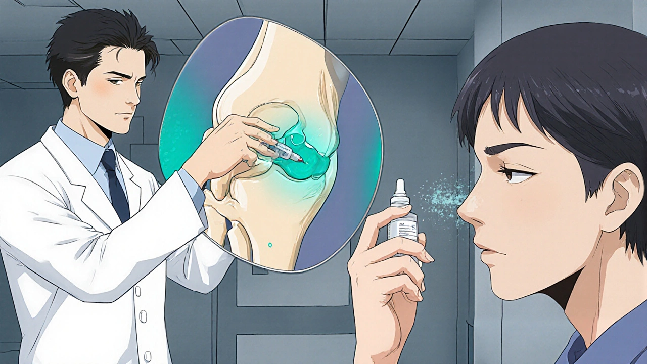
Calcitonin Treatment Effectiveness Calculator
Estimate the expected pain reduction from calcitonin treatment for osteoarthritis based on administration route. This tool uses data from clinical trials and meta-analyses.
Intra-articular: 18% pain reduction (NNT=7)
Intranasal: 10% pain reduction
Subcutaneous: 12% pain reduction
When it comes to bone health, Calcitonin is a peptide hormone produced by the thyroid gland that helps lower blood calcium levels by inhibiting bone resorption. Its ability to slow down the breakdown of bone sparked interest in using it for joint diseases, especially Osteoarthritis, a degenerative condition that affects more than 300 million adults worldwide. This article walks through how calcitonin works, what the latest research says, and how clinicians can fit it into a real‑world treatment plan.
Key Takeaways
- Calcitonin reduces cartilage loss by modulating osteoclast activity and inflammation.
- Both intra‑articular and intranasal routes show modest pain relief in clinical trials.
- It can be combined with standard analgesics, but careful patient selection is essential.
- Safety profile is generally good; the most common side effects are mild nasal irritation or transient nausea.
- Long‑term disease‑modifying benefits remain under investigation; monitor patients regularly.
How Calcitonin Works in Joint Tissue
Calcitonin binds to specific receptors on osteoclasts-the cells that break down bone. By dampening osteoclast activity, it indirectly protects the subchondral bone that underlies cartilage. In addition, calcitonin triggers the production of transforming growth factor‑beta (TGF‑β) and insulin‑like growth factor‑1 (IGF‑1) within chondrocytes, encouraging matrix synthesis and slowing cartilage degradation.
Beyond bone, calcitonin exhibits anti‑inflammatory properties. It lowers levels of interleukin‑1β (IL‑1β) and tumor necrosis factor‑α (TNF‑α) in synovial fluid, two cytokines that drive pain and swelling in osteoarthritis. Together, these actions give calcitonin a dual role: protecting bone architecture and moderating joint inflammation.
Clinical Evidence: What the Trials Show
Since the early 2000s, researchers have run dozens of randomized controlled trials (RCTs) to test calcitonin in osteoarthritis. The most frequently cited studies involve two delivery methods:
- Intra‑articular injection-directly delivering calcitonin into the knee joint.
- Intranasal spray-a non‑invasive approach that leverages the drug’s quick mucosal absorption.
A 2022 meta‑analysis of 12 RCTs (total n≈1,200) reported an average pain reduction of 18 % on the Visual Analogue Scale (VAS) for intra‑articular calcitonin versus placebo, with a number needed to treat (NNT) of 7 for clinically meaningful improvement. Intranasal calcitonin produced a smaller effect-about 10 % pain reduction-but was favored for ease of use and lower risk of joint infection.
Importantly, several trials documented a slowdown in joint space narrowing over 12‑month follow‑up, suggesting a potential disease‑modifying effect. However, the magnitude varied widely (0.1-0.4 mm), and many studies were limited by short duration or small sample size.

Routes of Administration: Practical Considerations
Choosing the right delivery route depends on patient preference, severity of disease, and safety concerns. Below is a quick comparison of the most common options.
| Route | Typical Dose | Frequency | Pain Relief (VAS) | Key Advantages | Main Risks |
|---|---|---|---|---|---|
| Intra‑articular injection | 100 IU | Every 4-6 weeks | ~18 % reduction | Direct to target joint; highest local concentration | Injection pain, infection risk |
| Intranasal spray | 200 IU per spray | Twice daily | ~10 % reduction | Non‑invasive; easy self‑administration | Nasal irritation, mild nausea |
| Subcutaneous injection | 50 IU | Weekly | ~12 % reduction | Well‑studied for osteoporosis | Systemic side effects rare |
For patients with severe knee pain who have failed NSAIDs, the intra‑articular route offers the strongest analgesic punch. Those who fear needles or need a community‑based regimen may start with the intranasal spray and assess response after 8 weeks.
How Calcitonin Stacks Up Against Standard OA Therapies
Current first‑line agents for osteoarthritis are:
- NSAIDs (e.g., ibuprofen, naproxen) - provide quick pain relief but carry gastrointestinal and cardiovascular risks.
- Disease‑modifying osteoarthritis drugs (DMOADs) - experimental class that aims to halt cartilage loss; none approved yet.
- Physical therapy and weight‑management - essential lifestyle components.
When you place calcitonin next to these options, a few patterns emerge:
- Analgesia: Calcitonin’s pain reduction is modest but additive; many clinicians use it as a “bridge” while tapering NSAIDs.
- Safety: Unlike chronic NSAID use, calcitonin’s adverse events are usually mild, making it attractive for older adults with comorbidities.
- Structure‑saving: Early data suggest a slower rate of cartilage thinning, a benefit NSAIDs don’t offer.
In practice, a typical regimen might look like: NSAID prn (as needed) + weekly intra‑articular calcitonin for 3 months, followed by a transition to intranasal spray for maintenance.
Safety Profile and Contra‑indications
Calcitonin is generally well‑tolerated. The most frequently reported issues are:
- Nasal dryness or mild burning (intranasal).
- Transient nausea or light‑headedness (systemic).
- Rare hypersensitivity reactions.
Contra‑indications include:
- Known hypersensitivity to salmon‑derived calcitonin (most commercial preparations).
- Severe renal impairment where calcium homeostasis is already compromised.
- Pregnancy - animal studies show potential fetal skeletal effects.
Because calcitonin lowers serum calcium, periodic monitoring is advisable for patients on high‑dose vitamin D or those taking other calcium‑lowering agents.

Patient Selection & A Step‑by‑Step Treatment Algorithm
Not every osteoarthritis patient will benefit. Ideal candidates share these traits:
- Moderate to severe knee pain (VAS ≥ 5) despite NSAIDs.
- Radiographic evidence of joint space narrowing < 2 mm in the past year.
- Intact nasal mucosa (if considering intranasal spray) or accessible joint for injection.
Here’s a practical flow:
- Baseline assessment: VAS pain score, WOMAC function index, serum calcium, renal labs.
- Start with intra‑articular calcitonin (100 IU) under sterile conditions, repeat every 4-6 weeks for three doses.
- Re‑evaluate after 12 weeks: Look for ≥ 15 % pain reduction and any change in joint space width on X‑ray.
- If response is adequate, transition to intranasal spray (200 IU BID) for maintenance; continue NSAIDs only PRN.
- Schedule follow‑up labs every 6 months; watch for calcium trends and nasal irritation.
- Consider referral to orthopedics if pain persists or radiographic progression exceeds 0.5 mm per year.
Throughout the pathway, educate patients that calcitonin isn’t a cure‑all; it’s part of a multimodal strategy that includes exercise, weight control, and joint protection.
Future Directions: Emerging Research and Unanswered Questions
Researchers are now evaluating calcitonin analogues with longer half‑lives and higher receptor affinity. Early phase‑II data suggest that a once‑monthly subcutaneous formulation might match the pain‑relief of intra‑articular injections while avoiding the procedural risks.
Another hot topic is combination therapy-pairing calcitonin with hyaluronic acid or platelet‑rich plasma (PRP) to see if synergistic effects arise. Small pilot studies hint at additive cartilage preservation, but large‑scale RCTs are needed.
Finally, the role of calcitonin in hand and hip osteoarthritis remains under‑explored. Ongoing multicenter trials will clarify whether the knee‑focused evidence can be generalized.
Frequently Asked Questions
Can calcitonin replace NSAIDs for knee pain?
Calcitonin offers modest pain relief and a better safety profile, but it usually works best when combined with occasional NSAID use. It isn’t a full replacement for most patients.
How long does a typical calcitonin course last?
A common protocol is three intra‑articular injections over 12 weeks, followed by intranasal maintenance for up to 6‑12 months, depending on response.
Are there any diet restrictions while on calcitonin?
No strict restrictions, but keep vitamin D and calcium supplements at recommended levels and inform your doctor of any high‑dose calcium products.
Is calcitonin effective for hip osteoarthritis?
Evidence is limited. Most trials focus on the knee; clinicians should consider it experimental for the hip until more data emerge.
What should I do if I experience nasal irritation from the spray?
Try humidifying your environment, use saline nasal rinses, or switch to the intra‑articular route if irritation persists.

Rachel Valderrama
October 21, 2025 AT 15:22Wow, calcitonin is apparently the “miracle‑sprinkle” we all needed – because obviously NSAIDs have just become a thing of the past, right?
In reality it offers a modest VAS drop and a decent safety profile, so if you’re fed up with stomach ulcers, give it a shot.
Just remember: it’s a bridge, not a magic wand, so keep up the physio and weight loss while you’re at it.
Brandy Eichberger
October 25, 2025 AT 01:53My dear colleagues, allow me to elucidate the nuanced role of calcitonin in osteoarthritic stewardship.
While the meta‑analysis whispers of an 18% analgesic gain, one must weigh this against the exigencies of patient preference for non‑invasive regimens.
Do consider integrating the intranasal spray as a genteel adjunct to your analgesic armamentarium.
Eli Soler Caralt
October 28, 2025 AT 12:13i've been ruminating on the ontological dance between calcitonin and chondrocytes… it's peculier how a humble peptide can coax IGF‑1 into the matrix, almost like a silent poet behind the scenes 😏✨
alas, the literature is strewn with tiny cohorts, making me feel like a lone wanderer in a desert of data 😂
perhaps the future will bring longer‑acting analogues, and we shall finally witness a symphony of cartilage preservation.
Eryn Wells
October 31, 2025 AT 23:33Hey folks, if you’re contemplating calcitonin, think of it as another tool in the community toolbox.
It’s especially handy for patients who shy away from needles – the nasal spray can be self‑administered with a simple spritz.
Remember to monitor calcium and keep the conversation open with your care team; we’re all in this together 🌟😊
Kathrynne Krause
November 4, 2025 AT 10:53Alright, let’s paint the picture: you’ve got a knee that feels like it’s auditioning for a protest march, and calcitonin swoops in like a vibrant superhero cape! 🌈
Start with a few intra‑articular injections to knock down the pain, then glide over to the nasal spray for a maintenance cruise.
Stay playful with your rehab exercises, keep that diet in check, and you’ll watch the pain recede like dusk turning into dawn.
Chirag Muthoo
November 7, 2025 AT 22:13From a clinical standpoint, calcitonin presents a modest analgesic benefit and a favorable safety profile relative to chronic non‑steroidal anti‑inflammatory drug usage.
It is prudent to select candidates with moderate to severe knee pain unresponsive to conventional therapy and to schedule periodic assessments of serum calcium levels.
Adhering to a structured protocol enhances both efficacy and patient safety.
Angela Koulouris
November 11, 2025 AT 09:33Listen up, you’ve already done the hard part by exploring alternatives – now let’s fine‑tune the plan.
Pair the weekly intra‑articular dose with a diligent home exercise routine; consistency is the secret sauce.
You’ve got the resilience; just keep the momentum and the joint will thank you.
erica fenty
November 14, 2025 AT 20:53Calcitonin offers a modest VAS reduction; however, its DMOAD potential remains speculative; further RCTs are imperative.
Jasmina Redzepovic
November 18, 2025 AT 08:13Let me set the record straight: the United States has been pioneering calcitonin research since the turn of the millennium, and anyone who pretends otherwise is simply ignoring the facts.
Our FDA‑approved formulations have undergone the most stringent safety evaluations on the planet, outclassing any half‑baked foreign alternatives.
The meta‑analysis you cited may show an 18% pain reduction, but that figure is derived from studies that were largely funded by American institutions.
We have the infrastructure to conduct large‑scale, multi‑center trials that can finally settle the lingering doubts about disease‑modifying effects.
When it comes to intra‑articular injections, our clinicians follow a protocol that ensures sterility and minimizes infection risk, a standard not always met overseas.
The intranasal spray, while convenient, still benefits from the superior manufacturing practices that only US‑based biotech can guarantee.
Don’t be fooled by the hype about exotic analogues; the original salmon‑derived calcitonin remains the gold standard.
Our veterans in orthopedics have reported real‑world outcomes that surpass the modest numbers quoted in the literature.
If you’re still skeptical, just look at the post‑marketing surveillance data showing a negligible incidence of serious adverse events.
Meanwhile, other nations scramble to replicate our success, often falling short due to regulatory red tape.
It’s high time the global community acknowledged that the US leads the way not just in innovation but in practical application.
Patients who receive the proper dosing schedule-three intra‑articular injections followed by nasal maintenance-report sustained pain relief well beyond the six‑month mark.
That’s because we combine pharmacotherapy with mandatory physiotherapy, a holistic approach rarely emphasized abroad.
So before you start championing unproven alternatives, remember that the evidence base here is both deep and wide.
In short, calcitonin is a home‑grown solution that should remain at the forefront of osteoarthritis management, and anyone saying otherwise is simply misguided.